Wednesday 19th of March 2025
Vet Advice

Tendon and Ligament Injuries

Scott Dunn’s Equine Clinic have put together this useful article looking at the different tendon and ligament injuries, the early signs, diagnosis and various different treatment options available to aid recovery.

What are tendons?

Tendons are the elastic structures that attach muscles to bones. Most tendons are relatively short and rarely damaged. However, the long tendons of the limbs are vulnerable to damage during exercise or as a result of trauma. The flexor tendons are the most important structures as discussed below.

tendon

Image showing location of tendons below the knee

Location of these tendons

The deep digital flexor tendon (DDFT) and the superficial digital flexor tendon (SDFT) run down the back of the limb from the level of the knee/hock.

The SDFT ends on the pastern and the DDFT ends on the back of the pedal bone. Around the knee, hock, fetlock and pastern regions the tendons are enclosed by a fluid filled sheath. Several strong, short, annular ligaments help to keep the tendons in place in regions of high movement such as joints. The tendons themselves are composed of longitudinally arranged bundles of fibres. Blood supply to tendons and ligaments is poor compared to muscles and other tissues.

bowed tendon

Image revealing a bowed tendon

The different types of tendon injuries

Injury tendons commonly occurs during exercise. Strenuous exercise can result in tearing of fibres especially in unfit horses. Even fit horses which are over stretching tendons in fast work or on un-level ground or during jumping at speed can damage these structures. The degree of damage can range from minor, with minimal fibre damage to severe with total tendon rupture.

A knock to a tendon may result in slight bruising or severe damage leading to tendon rupture. Sharp trauma which cuts through the skin can vary from minor tendon damage to partial or full thickness laceration of the tendon. If a tendon sheath is involved these can lead to potentially life threatening infection if not dealt with promptly.

First signs of tendon injury

Damage to a tendon usually results in inflammation which we commonly feel as heat and swelling. Minor fibre damage leads to slight enlargement of the affected part of the tendon which feels warmer than the corresponding area of the opposite limb. Mild sprains often do not cause lameness. If there is severe damage, the limb can become very painful, with the toe tipped upwards or the fetlock may sink at the walk. In cases of tendon sheath sepsis the horse will also be very lame.

Ultrasound scan revealing a core lesion of the SDFT in the left image. The right image shows the lack of fibre pattern on a longitudinal scan of the SDFT at the top of the image

Ultrasound scan revealing a core lesion of the SDFT in the left image. The right image shows the lack of fibre pattern on a longitudinal scan of the SDFT at the top of the image

Diagnosis of tendon injuries

If you suspect that your horse has a tendon injury, you should call the clinic for advice and an appointment. It is difficult to accurately assess the extent of the damage by look and palpation alone, so an ultrasound scan, approximately one week after injury will allow us to visualise the damaged structure(s), if it is above the hoof capsule.

Treatment options

There are several different treatment options for tendon injuries.

Initial treatment in the 10-14 days after an injury usually involves;

·    Box rest
·    Ice application or cold hosing 2-3 times daily and/or application of kaolin poultice
·    Bandaging to immobilise the limb
·    Anti-inflammatories such as bute to aid in reduction of swelling and provide pain relief.

These steps are aimed at reducing the initial inflammation and pain along with preventing any further injury. Once the initial inflammation has stabilised a controlled exercise programme can be started.

Controlled Exercise

This is the most important aspect of treatment. This will generally start with hand walking while still on box rest with gradual increasments every 1-2 weeks for a period of 3 months. In certain cases an initial period of total box rest is advised. Horses are often out of work for 6-12 months with these injuries. Controlled exercise helps the new tendon fibres align longitudinally ultimately resulting in increased strength and flexibility.

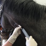
Repeat ultrasound scans are invaluable as an aid to determine the healing process and adjust the exercise programme accordingly, such as when to introduce trot work or steady canter exercise.

Other treatments options

None of the treatments available reduce the horse’s lay off period. But the aim is to improve the quality of repair and reduce the risk of re-injury on return to exercise.

Tendon splitting or fenestration. A scalpel incision or lots of needle holes can be made to release the initial blood clot from a core lesion and this may help new blood vessels to grow into the injured area. This treatment requires to be done within 1-2 weeks of the initial injury,

·   Tendon injections.  A group of drugs called PSGAG’s (Adequan / Cartrophen) can be used successfully to inject tendon injuries and assists in short term healing. More commonly we now use either stem cells or platelet rich plasma, both of which are injected directly into the tendon soon after injury.

·   Surgery.  This is sometimes performed in the case of tendon lacerations to help oppose the edges of the damaged tendon or in the case of tendon sheath sepsis to remove infection both of these procedures are carried out under general anaesthesia.

Ligament injuries

Ligaments are the elastic soft tissue structures that connect the ends of bone at joints. In certain cases they attach from a bone to a tendon e.g. the inferior check ligament. Their role is to maintain bones in alignment and provide support to a joint. They are usually located on either side of a joint. Therefore the name collateral is often in their name relating to positioning. Some joints have one ligament on each side and others such as the stifle.

Ultrasound scan of medial meniscus of stifle revealing irregular medial meniscus

Ultrasound scan of medial meniscus of stifle revealing irregular medial meniscus

How are ligament injuries caused?

They can occur in multiple ways such as direct trauma, abnormal or excessive forces placed on a joint eg. turning at speed. In cases of complete rupture there can be instability of the affected joint. Common Ligament injuries in the horse include:

·      Suspensory ligament desmitis

·      Collateral ligaments of the coffin joint, fetlock joint and hock joints

·      Palmar annular ligament of the fetlock

·      Accessory (check) ligament of the deep flexor tendon

·      Meniscal and cruciate ligaments of the stifle.

Diagnosis of ligament injuries

In severe cases of ligament injury the presence of heat, swelling and pain on palpation may be present.  To aid localisation of injury, ultrasound can confirm the diagnosis. In other cases which are more subtle or involved within the hoof a full lameness, further investigation may be required. This would involve such procedures as nerve blocks followed by radiography, ultrasonography and in certain cases advanced procedures such as magnetic resonance imaging (MRI) to obtain a diagnosis.

Treatment of ligament injuries

The initial treatment of these injuries are often managed similar to that of tendon injuries and can be a combination of:

·    Box rest.
·    Ice application or cold hosing 2-3 times daily and/or application of kaolin poultice
·    Bandaging to immobilise the limb
·    Anti-inflammatories such as ‘bute’ to aid in reduction of swelling and provide pain relief.

This is often followed by a slow rehabilitation plan which often takes at least 9 month to resume normal exercise programme if successful. This rehabilitation might initially involve a period of box rest followed by controlled walking, often for 3 months.

Portable shockwave machine

Portable shockwave machine

Other treatment options

These treatment options are similar to those of tendon injuries:

·      Surgery can be used to evaluate further the extent of injury or debride lesions. In proximal suspensory desmitis a lateral plantar nerve neurectomy and fasciotomy is sometimes performed to alleviate symptoms of lameness.

·      Platelet derived plasma

·      Stem cells

·      Extracorporal shockwave therapy. This involves the delivery of high impact short duration physical ‘shock waves’ to an area of damaged or inflamed tissue.

www.scott-dunns.co.uk

You may also like…

Join us on Facebook


Follow on Twitter